
TRACK 1: Inspiring Innovation in Formulation, Bioprocessing, and Drug Delivery
Category: Poster Abstract

Leah Schrass
Research Assistant
University of Kentucky
Lexington, Kentucky, United States
Leah Schrass
Research Assistant
University of Kentucky
Lexington, Kentucky, United States
Daniel Kolpek
University of Kentucky, United States
Daniel Pack
University of Kentucky, United States
Jonghyuck Park
University of Kentucky, United States
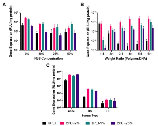 Figure 1: Changes in Transfection Efficiency with Serum Exposure Transfection efficiency was measured in HeLa cells using a range of zPEI nanoparticles after serum exposure compared to uPEI nanoparticles. A) Analysis of the effects of increasing fetal bovine serum (FBS) concentration on transfection efficiency with pre-incubation of nanoparticles before introduction to cell culture. B) Evaluation of the effects of weight ratio on transfection efficiency for zPEI and uPEI nanoparticles pre-incubated with 50% FBS. C) Determination of the transfection efficiency for zPEI nanoparticles pre-incubated in 50% human serum (HS) and human plasma (HP). All error bars represent standard deviation.
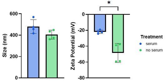 Figure 2: Size and Charge Differences with PLGA-PEMA Nanoparticle Serum Exposure Size and zeta potential were measured after PLGA-PEMA exposure to 50% FBS. The size of the nanoparticles increased by ~80 nm and the charge decreased ~ 30 mV after protein corona formation. Error bars represent standard deviation and * is a p-value < 0.05.
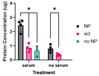 Figure 3: PLGA-PEMA Protein Corona Concentration after Centrifugation Purification The concentration of protein in the PLGA-PEMA nanoparticles was evaluated after exposure to 50% FBS and a three-wash centrifugation procedure (16,000 g for 10 min). Final concentration of protein in the corona (NP) was greater than the concentration in the third wash solution (w3) and a centrifuged solution of FBS alone (no NP). Error bars represent standard deviation and * is a p-value < 0.05.