
TRACK 1: Inspiring Innovation in Formulation, Bioprocessing, and Drug Delivery
Category: Poster Abstract

Austin Daniels, Ph.D.
Application Scientist
Yokogawa Fluid Imaging Technologies
Scarborough, Maine, United States
Austin Daniels, Ph.D.
Application Scientist
Yokogawa Fluid Imaging Technologies
Scarborough, Maine, United States
Sigrid Kuebler, Ph.D.
Marketing Director
Yokogawa Fluid Imaging Technologies
Scarborough, Maine, United States
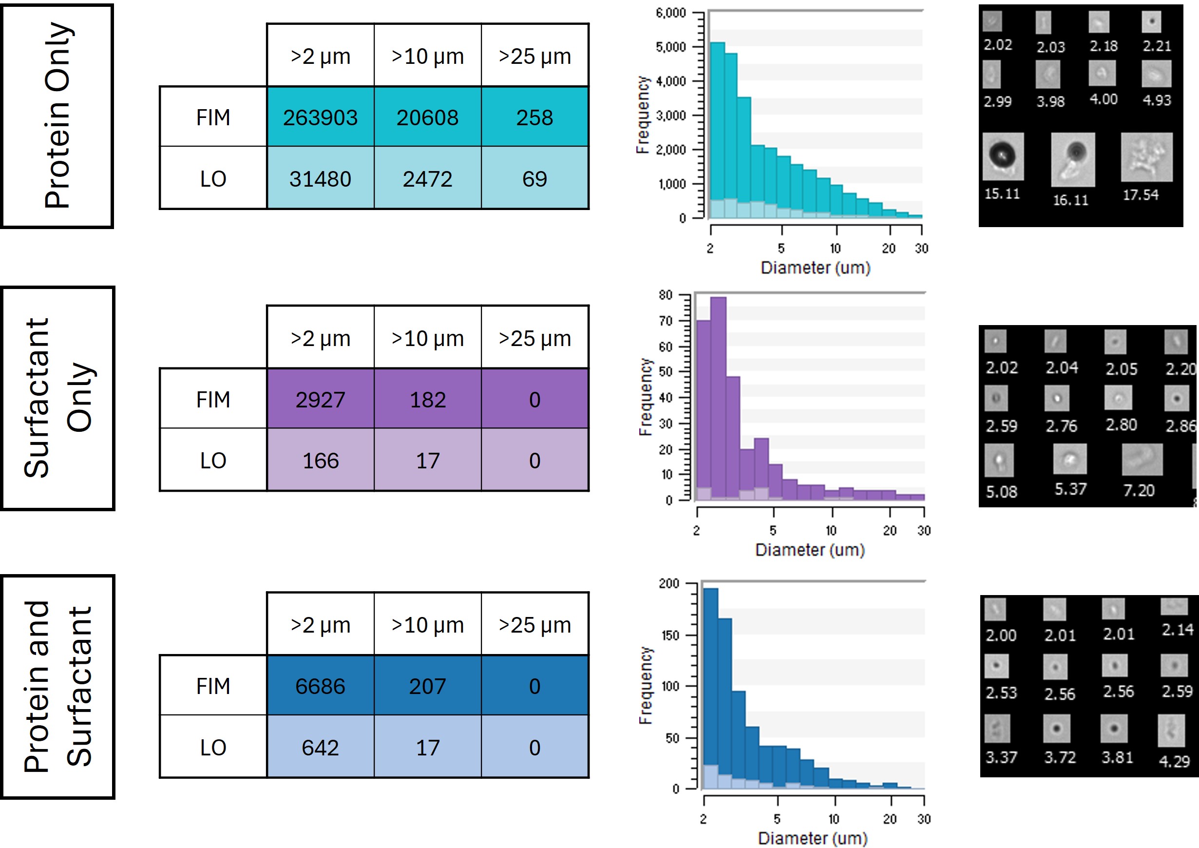 Subvisible particles measured in different formulations following agitation stress. Dark shaded regions indicate FIM measurements and light shaded regions indicate LO measurements. (Left) particle counts measured in different size ranges. (Center) Particle size distributions measured for each sample. (Right) Collages of sample particle images captured by FIM.
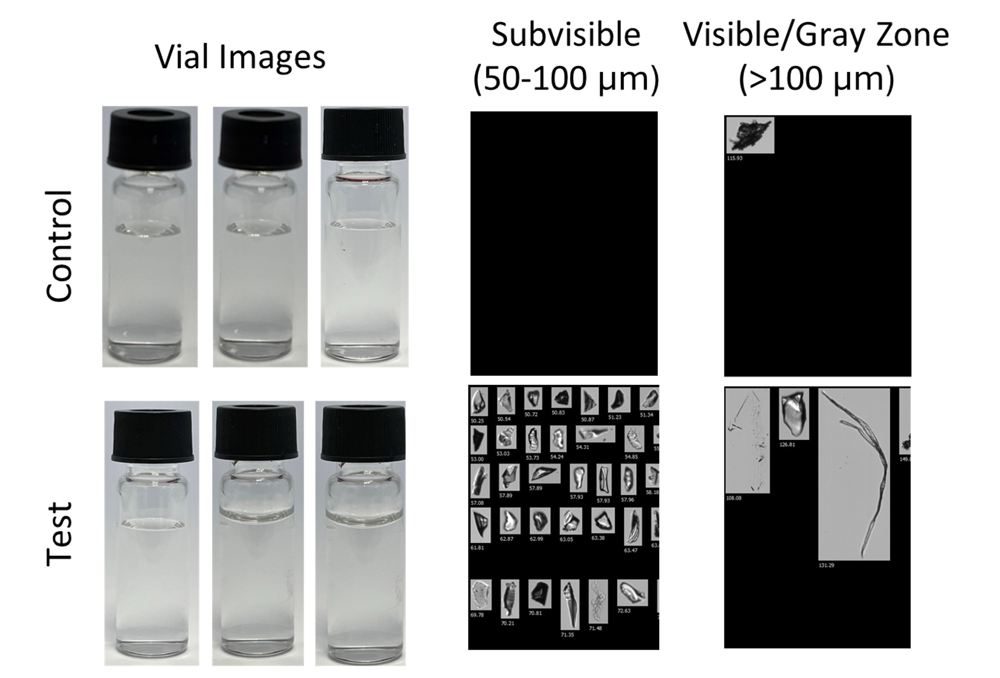 (Left) Images of control and test vials taken inside a light box. No visible particles were observed while inspecting the particles in the lightbox. (Right) collages of particles observed via FIM in the control and test vials. Collages on the left contain subvisible particles 50-100 μm in size, while the collage on the right contain visible and gray zone particles >100 μm in size.
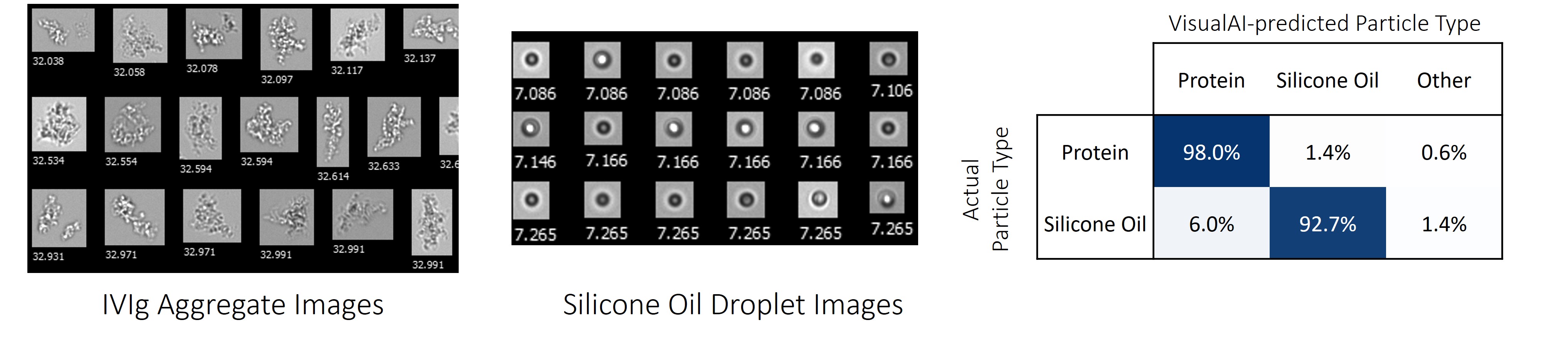 (Left and center) Collages of sample FIM images of protein aggregates and silicone oil droplets. (Right) confusion matrix showing the classification performance of VisualAI on FIM images known to contain a protein aggregate or silicone oil droplet.